


Sulfur is called nature's "beauty mineral" because it keeps skin smooth and youthful and hair glossy. Sulfur is necessary for the production of collagen and keratin, proteins necessary for the health and maintenance of skin, nails and hair'
$14.95
|
|
Imaging Services >
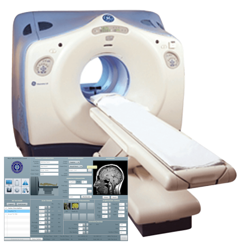
CT Scan
Computed Tomography (CT Scan)
For an appointment for CT Scan - Click HERE What can a person expect during a CT procedure? How is CT used in cancer screening? What is total, or whole-body, CT? Is the radiation from CT harmful? What are the risks of CT scans for children? What is computed tomography? Computed tomography (CT) is an imaging procedure that uses special x-ray equipment to create detailed pictures, or scans, of areas inside the body. It is also called computerized tomography and computerized axial tomography (CAT). The term tomography comes from the Greek words tomos (a cut, a slice, or a section) and graphein (to write or record). Each picture created during a CT procedure shows the organs, bones, and other tissues in a thin “slice” of the body. The entire series of pictures produced in CT is like a loaf of sliced bread—you can look at each slice individually (2-dimensional pictures), or you can look at the whole loaf (a 3-dimensional picture). Computer programs are used to create both types of pictures. Most modern CT machines take continuous pictures in a helical (or spiral) fashion rather than taking a series of pictures of individual slices of the body, as the original CT machines did. Helical CT has several advantages over older CT techniques: it is faster, produces better 3-D pictures of areas inside the body, and may detect small abnormalities better. The newest CT scanners, called multislice CT or multidetector CT scanners, allow more slices to be imaged in a shorter period of time. In addition to its use in cancer, CT is widely used to help diagnose circulatory (blood) system diseases and conditions, such as coronary artery disease (atherosclerosis), blood vessel aneurysms, and blood clots; spinal conditions; kidney and bladder stones; abscesses; inflammatory diseases, such as ulcerative colitis and sinusitis; and injuries to the head, skeletal system, and internal organs. CT can be a life-saving tool for diagnosing illness and injury in both children and adults. What can a person expect during a CT procedure? During a CT procedure, the person lies very still on a table, and the table passes slowly through the center of a large x-ray machine. With some types of CT scanners, the table stays still and the machine moves around the person. The person might hear whirring sounds during the procedure. At times during a CT procedure, the person may be asked to hold their breath to prevent blurring of the images. Sometimes, CT involves the use of a contrast (imaging) agent, or “dye.” The dye may be given by mouth, injected into a vein, given by enema, or given in all three ways before the procedure. The contrast dye highlights specific areas inside the body, resulting in clearer pictures. Iodine and barium are two dyes commonly used in CT. In very rare cases, the contrast agents used in CT can cause allergic reactions. Some people experience mild itching or hives (small bumps on the skin). Symptoms of a more serious allergic reaction include shortness of breath and swelling of the throat or other parts of the body. People should tell the technologist immediately if they experience any of these symptoms, so they can be treated promptly. Very rarely, the contrast agents used in CT can also cause kidney problems in certain patients. These kidney problems usually do not have any symptoms, but they can be detected by running a simple test on a blood sample. CT does not cause any pain. However, lying in one position during the procedure may be slightly uncomfortable. The length of a CT procedure depends on the size of the area being scanned, but it usually lasts only a few minutes to half an hour. For most people, the CT is performed on an outpatient basis at a hospital or a radiology center, without an overnight hospital stay. Some people are concerned about experiencing claustrophobia during a CT procedure. However, most CT scanners surround only portions of the body, not the whole body. Therefore, people are not enclosed in a machine and are unlikely to feel claustrophobic. Women should let their health care provider and the technologist know if there is any possibility that they are pregnant, because radiation from CT can harm a growing fetus. Patient undergoing CT of the abdomen. Drawing shows the patient on a table that slides through the CT machine, which takes x-ray pictures of the inside of the body. How is CT used in cancer? CT is used in cancer in many different ways:
How is CT used in cancer screening? Studies have shown that CT can be effective in both colorectal cancer screening (including screening for large polyps) and lung cancer screening. Colorectal cancer CT colonography (also known as virtual colonoscopy) can be used to screen for both large colorectal polyps and colorectal tumors. CT colonography uses the same dose of radiationthat is used in standard CT of the abdomen and pelvis, which is about 10 millisieverts (mSv) (1). (By comparison, the estimated average annual dose received from natural sources of radiation is about 3 mSv.) As with standard (optical) colonoscopy, a thorough cleansing of the colon is performed before this test. During the examination, air or carbon dioxide gas is pumped into the colon to expand it for better viewing. The National CT Colonography Trial, an NCI-sponsored clinical trial, found that the accuracy of CT colonography is similar to that of standard colonoscopy. CT colonography is less invasive than standard colonoscopy and has a lower risk of complications. However, if polyps or other abnormal growths are found on CT colonography, a standard colonoscopy is usually performed to remove them. Whether CT colonography can help reduce the death rate from colorectal cancer is not yet known, and most insurance companies (and Medicare) do not currently reimburse the costs of this procedure. Also, because CT colonography can produce images of organs and tissues outside the colon, it is possible that non-colorectal abnormalities may be found. Some of these "extra-colonic" findings will be serious, but many will not be, leading to unnecessary additional tests and surgeries. Lung cancer The NCI-sponsored National Lung Screening Trial (NLST) showed that people aged 55 to 74 years with a history of heavy smoking are 20 percent less likely to die from lung cancer if they are screened with low-dose helical CT than if they are screened with standard chest x-rays. (Previous studies had shown that screening with standard chest x-rays does not reduce the death rate from lung cancer.) The estimated amount of radiation in a low-dose helical CT procedure is 1.5 mSv (1). Despite the effectiveness of low-dose helical CT for lung cancer screening in heavy smokers, the NLST identified risks as well as benefits. For example, people screened with low-dose helical CT had a higher overall rate of false-positive results (that is, findings that appeared to be abnormal even though no cancer was present), a higher rate of false-positive results that led to an invasive procedure (such as bronchoscopy or biopsy), and a higher rate of serious complications from an invasive procedure than those screened with standard x-rays. NCI’s Patient and Physician Guide: National Lung Screening Trial provides more information on the benefits and harms. The benefits of helical CT in screening for lung cancer may vary, depending on how similar someone is to the people who participated in the NLST. The benefits may also be greater for those with a higher lung cancer risk, and the harms may be more pronounced for those who have more medical problems (like heart or other lung disease), which could increase problems arising from biopsies and other surgery. People who think that they have an increased risk of lung cancer and are interested in screening with low-dose helical CT should discuss the appropriateness and the benefits and risks of lung cancer screening with their doctors. They should also be aware that, because the technique is fairly new, some insurance plans do not currently cover it. What is total, or whole-body, CT? Total, or whole-body, CT creates pictures of nearly every area of the body—from the chin to below the hips. This procedure, which is used routinely in patients who already have cancer, can also be used in people who do not have any symptoms of disease. However, whole-body CT has not been shown to be an effective screening method for healthy people. Most abnormal findings from this procedure do not indicate a serious health problem, but the tests that must be done to follow up and rule out a problem can be expensive, inconvenient, and uncomfortable. In addition, whole-body CT can expose people to relatively large amounts of ionizing radiation—about 12 mSv, or four times the estimated average annual dose received from natural sources of radiation. Most doctors recommend against whole-body CT for people without any signs or symptoms of disease. What is combined PET/CT? Combined PET/CT uses two imaging methods, CT and positron emission tomography(PET), in one procedure. CT is done first to create anatomic pictures of the organs and structures in the body, and then PET is done to create colored pictures that show chemical or other functional changes in tissues. Different types of positron-emitting (radioactive) substances can be used in PET. Depending on the substance used, different kinds of chemical or functional changes can be imaged. The most common type of PET procedure uses an imaging agent called FDG (a radioactive form of the sugar glucose), which shows the metabolic activity of tissues. Because cancerous tumors are usually more metabolically active than normal tissues, they appear different from other tissues on a PET scan. Other PET imaging agents can provide information about the level of oxygen in a particular tissue, the formation of new blood vessels, the presence of bone growth, or whether tumor cells are actively dividing and growing. Combining CT and PET may provide a more complete picture of a tumor’s location and growth or spread than either test alone. The combined procedure may improve the ability to diagnose cancer, to determine how far a tumor has spread, to plan treatment, and to monitor response to treatment. Combined PET/CT may also reduce the number of additional imaging tests and other procedures a patient needs. Is the radiation from CT harmful? Some people may be concerned about the amount of radiation they receive during CT. CT imaging involves the use of x-rays, which are a form of ionizing radiation. Exposure to ionizing radiation is known to increase the risk of cancer. Standard x-ray procedures, such as routine chest x-rays and mammography, use relatively low levels of ionizing radiation. The radiation exposure from CT is higher than that from standard x-ray procedures, but the increase in cancer risk from one CT scan is still small. Not having the procedure can be much more risky than having it, especially if CT is being used to diagnose cancer or another serious condition in someone who has signs or symptoms of disease. It is commonly thought that the extra risk of any one person developing a fatal cancer from a typical CT procedure is about 1 in 2,000 (2). In contrast, the lifetime risk of dying from cancer in the U.S. population is about 1 in 5 (3). It is also important to note that everyone is exposed to some background level of naturally occurring ionizing radiation every day. The average person in the United States receives an estimated effective dose of about 3 millisieverts (mSv) per year from naturally occurring radioactive materials, such as radon and radiation from outer space (1). By comparison, the radiation exposure from one low-dose CT scan of the chest (1.5 mSv) is comparable to 6 months of natural background radiation, and a regular-dose CT scan of the chest (7 mSv) is comparable to 2 years of natural background radiation (1). The widespread use of CT and other procedures that use ionizing radiation to create images of the body has raised concerns that even small increases in cancer risk could lead to large numbers of future cancers (4, 5). People who have CT procedures as children may be at higher risk because children are more sensitive to radiation and have a longer life expectancy than adults. Women are at a somewhat higher risk than men of developing cancer after receiving the same radiation exposures at the same ages (6). People considering CT should talk with their doctors about whether the procedure is necessary for them and about its risks and benefits. Some organizations recommend that people keep a record of the imaging examinations they have received in case their doctors don’t have access to all of their health records. A sample form, called My Medical Imaging History, was developed by the Radiological Society of North America, the American College of Radiology, and the U.S. Food and Drug Administration. It includes questions to ask the doctor before undergoing any x-ray exam or treatment procedure. What are the risks of CT scans for children? Radiation exposure from CT scans affects adults and children differently. Children are considerably more sensitive to radiation than adults because of their growing bodies and the rapid pace at which the cells in their bodies divide. In addition, children have a longer life expectancy than adults, providing a larger window of opportunity for radiation-related cancers to develop (7). Individuals who have had multiple CT scans before the age of 15 were found to have an increased risk of developing leukemia, brain tumors (8), and other cancers (9) in the decade following their first scan. However, the lifetime risk of cancer from a single CT scan was small—about one case of cancer for every 10,000 scans performed on children. In talking with health care providers, three key questions that the parents can ask are: why is the test needed? Will the results change the treatment decisions? Is there an alternative test that doesn’t involve radiation? If the test is clinically justified, then the parents can be reassured that the benefits will outweigh the small long-term risks. Selected References
|
|
|

OPEN 24 HOURS: ACCIDENT EMERGENCY, LAB SERVICES, IMAGING SERVICES & PHARMACY


Medical Disclaimer: The Contents of this site is not intended to be a substitute for professional medical advice, diagnosis, or treatment. Always seek the advice of your physician or other qualified health provider with any questions you may have regarding a medical condition or treatment.


Medical Disclaimer: The Contents of this site is not intended to be a substitute for professional medical advice, diagnosis, or treatment. Always seek the advice of your physician or other qualified health provider with any questions you may have regarding a medical condition or treatment.