|
Magnesium has an effect on more than 300 bodily chemical reactions. This includes maintaining heart health, sustaining blood vessels, boosting and maintaining energy levels, producing new cells and proteins, and enabling enzyme activity.
|
|
Imaging Services
Imaging ServicesOur team focuses on providing patients with the highest quality medical imaging services in Nigeria. We know that getting the best possible image of an injury or condition within your body is incredibly important for diagnosis and a successful medical treatment. Our medical imaging service are reports from staff of board certified radiologists licensed technologists are fully accredited by the American College of Radiology. Your health and safety are our top priority. We use the most technologically sophisticated imaging technology available. The advanced medical imaging technology we employ includes: Computed Tomography (CT)
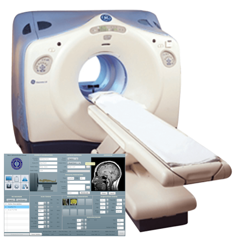
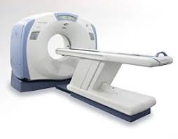
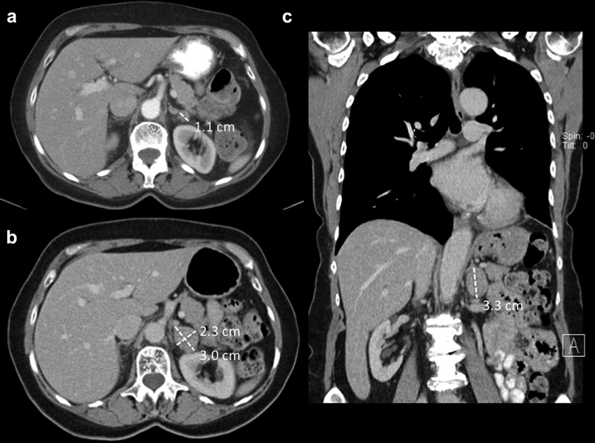
One of the most modern of diagnostic imaging techniques, Computed Tomography (CT) – popularly known as a CAT scan – combines multiple X-ray projections from different angles to create images. The CT procedure employs a series of X-ray beams that produce a cross-section of images the Computed Tomography scanner constructs into detailed 3-D pictures. CT can diagnose medical abnormalities such as tumors, cysts, bone calcification, internal bleeding and many different types of cancer. CT scans are commonly used to evaluate the following:
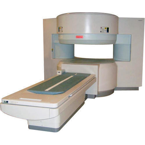
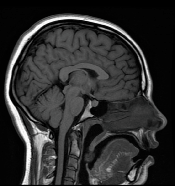
Magnetic Resonance Imaging (MRI)Magnetic Resonance Imaging (MRI) is a medical imaging procedure that employs radio waves and a magnetic field to produce detailed pictures of internal body structures, including organs and tissue. MRI is a highly effective tool in diagnosing a wide range of medical conditions such as tumors, orthopedic injuries, neurological problems and other medical issues. MRI imaging does not use radiation to produce the image. When receiving an MRI, a patient lies comfortably on a bed which is then inserted into an MRI scanner. The scanner is a large circular magnet that creates a magnetic field strong enough to align protons in hydrogen atoms inside a patient’s body. Then, patients are exposed to a radio wave. Much like an ultrasound, the radio wave bounces back and is interpreted by the MRI scanner as an image. MRI is often used to evaluate the following:
X-RayDiscovered over 100 years ago, the most commonly used form of medical imaging is X-ray technology. X-rays utilize ionizing radiation to create images of structures inside the body. This is accomplished through the emission of X-ray beams into the body which are absorbed in differently by the varied densities of material inside the body. For example, in an X-ray a bone or a foreign object inside your body will be visible, but muscle or tissue is not. X-rays are used to diagnose a wide variety of medical conditions. X-rays are typically used to evaluate the following:
UltrasoundOtherwise known as medical sonography, diagnostic ultrasound employs high frequency sound waves to provide images of the body. An ultrasound works by sending sound waves directly into the body. When these sound waves bounce back, the ultrasound machine interprets these sound waves into an image. Unlike an X-ray, an ultrasound does not use radiation to produce a diagnostic image. This makes ultrasound medical imaging not only painless, but safe. Ultrasound is commonly used to evaluate the following:
Magnetic resonance imaging (MRI) of the breast uses a powerful magnetic field, radio waves and a computer to produce detailed pictures of the structures within the breast.
Magnetic resonance imaging (MRI) of the prostate uses a powerful magnetic field, radio waves and a computer to produce detailed pictures of the structures within a man's prostate gland.
Computed tomography (CT) is an imaging procedure that uses special x-ray equipment to create detailed pictures, or scans, of areas inside the body.
Sonography or ultrasound tests use sound waves to create images of what’s happening inside the body.
Acronym for Computed (Axial) Tomography and Magnetic Resonance Imaging.MRI machines do not emit ionizing radiation.Suited for bone injuries, Lung and Chest imaging, cancer detection.
X-ray image is produced when a small amount of radiation is passed through a body part to create an image. The image is produced as a result of the ways in which different internal structures absorb the radiation.
|
|
|

OPEN 24 HOURS: ACCIDENT EMERGENCY, LAB SERVICES, IMAGING SERVICES & PHARMACY


